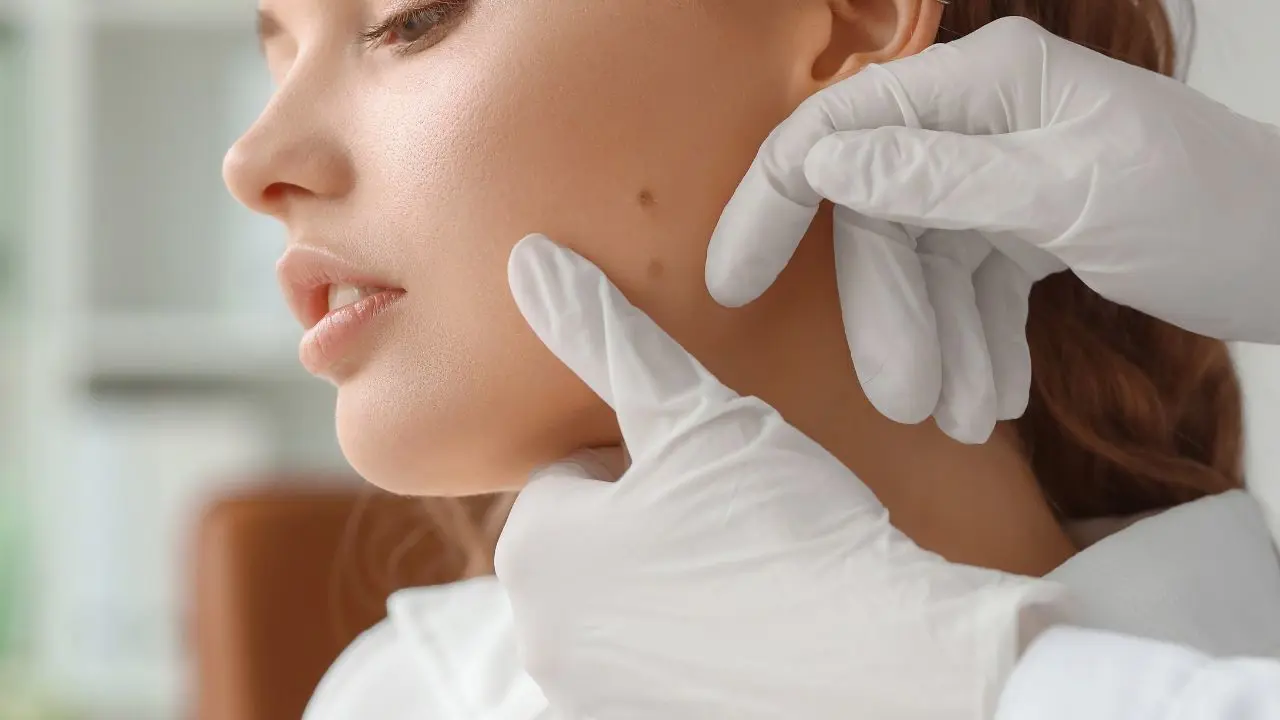
Assessing moles and considering their removal is a common practice in dermatology, particularly when there are concerns about changes in appearance, size, color, or symmetry, which could indicate potential skin cancer or other issues. Mole assessment and removal are common practices in dermatology aimed at evaluating moles for signs of skin cancer or other concerning changes and, if necessary, removing them for diagnostic or cosmetic purposes.
Mole Assessment
Self-Examination: Regular self-examination of moles is essential. Look for the ABCDE signs of melanoma:
- Asymmetry: One half of the mole does not match the other half.
- Border: The edges are irregular, ragged, blurred, or notched.
- Color: The color is not uniform, with shades of brown, black, blue, red, white, or pink.
- Diameter: The diameter is larger than 6 millimeters (about the size of a pencil eraser) or is increasing in size.
- Evolution: The mole is changing in size, shape, color, or elevation, or new symptoms such as itching, bleeding, or crusting develop.
Dermatological Evaluation: If you notice any concerning changes in a mole or have a personal or family history of skin cancer, consult a dermatologist for a comprehensive evaluation.
Dermoscopy: Dermoscopy is a non-invasive technique that allows dermatologists to examine moles under magnification and assess their structural features, helping to differentiate between benign and malignant lesions.
Biopsy: If a mole appears suspicious, a dermatologist may perform a biopsy, in which a sample of tissue is removed and examined under a microscope to determine if cancerous cells are present.
Mole Removal:
Indications: Mole removal may be recommended for cosmetic reasons or if the mole is suspicious for skin cancer or causing symptoms such as itching, irritation, or bleeding.
Methods:
- Excisional Biopsy: The mole is surgically removed along with a margin of surrounding normal tissue. This method ensures complete removal and allows for further examination under a microscope.
- Shave Biopsy: The mole is shaved off at the surface of the skin using a scalpel or blade. This method is less invasive but may not provide a complete sample for examination.
- Laser Removal: Lasers can be used to target and destroy pigment cells in the mole, causing it to fade or disappear gradually. This method is often used for small, non-cancerous moles and may require multiple sessions.
Post-Removal Care: After mole removal, it's essential to follow your dermatologist's post-procedure instructions, which may include keeping the area clean and dry, avoiding sun exposure, and applying topical medications as prescribed.
Pathology Examination: If a mole is removed due to suspicion of skin cancer, the tissue sample will be sent to a laboratory for further examination by a pathologist to confirm the diagnosis and assess the margins for any remaining cancer cells.
Follow-Up: Regular follow-up appointments with your dermatologist are important to monitor healing, check for signs of recurrence, and assess for the development of new moles or skin lesions.
Overall, mole assessment and removal should be approached with caution and performed by qualified dermatologists to ensure proper diagnosis, treatment, and monitoring. If you have any concerns about a mole or skin lesion, seek medical advice promptly.
